
Do I Have a Herniated Disk? Signs That Matter
Back pain alone can't confirm a herniated disk; pain traveling into an arm or leg, numbness, or weakness on one side is more suggestive. An MRI finding without a matching symptom pattern and examination isn't proof that the disk is causing your pain.
You lift a laundry basket, straighten up, and feel a sharp catch in your lower back. Then you notice a line of pain down your leg, and suddenly you're wondering whether something has slipped out of place.
The useful question isn't just how much it hurts. It's where the symptoms travel, whether strength or sensation has changed, and whether anything has changed with bladder, bowel, or saddle-area feeling.
Pain Patterns That Point Toward a Herniated Disk
One-sided pain that travels away from the spine is a more useful clue than the location of the backache itself. A lower-back problem may produce pain through the buttock and leg, while a neck problem may affect the shoulder, arm, or hand.
A herniated disk occurs when soft inner material pushes through a tear in its tougher outer layer. If that material irritates a nearby nerve, symptoms can include radiating pain, tingling, numbness, or weakness, as described in Mayo Clinic's herniated disk guidance.
Compare the pattern, not just the sore spot
Use this table to organize what you notice. It's a symptom map to discuss with a clinician, not a way to confirm the diagnosis at home.
| What you notice | Leans toward disk-related nerve irritation | More often something else |
|---|---|---|
| Pain stays in the low back or neck | Possible, but not specific | Muscle strain, joint irritation, or arthritis |
| Pain travels down one leg past the knee, or into one arm and hand | Yes, a typical radiating nerve pattern | Less typical of a simple strain |
| Numbness or tingling in a strip of skin | Yes, suggests nerve involvement | A diffuse ache is less specific |
| New weakness on one side, such as foot drag or weak grip | Yes, needs a prompt examination | Fatigue or soreness alone doesn't establish nerve weakness |
| Pain jumps with a cough, sneeze, or strain | Supports nerve irritation | This response isn't required for the diagnosis |
| Stiffness worst in the morning that eases as you move | Not a strong disk clue by itself | Can occur with arthritis or other causes of stiffness |
| A localized sore muscle that's tender to press and settles with reduced loading | Less suggestive without radiating or neurologic symptoms | Muscle strain is another possibility |
These patterns overlap. Morning stiffness doesn't prove arthritis, and tenderness doesn't rule out a disk problem. A clinician looks for a combination of findings rather than assigning a diagnosis from a single row.
Notice what the limb can do
Weakness means more than feeling reluctant to move because it hurts. Concrete examples include a foot catching on the floor, trouble pushing up onto the toes, or objects slipping from your hand.
Don't repeatedly test a painful limb to exhaustion. Write down what happened during ordinary activities and whether it's becoming more noticeable.
For lower-back symptoms, understanding what causes sciatica nerve pain can help you describe the route into your leg. Sciatica describes a nerve-related symptom pattern, not automatic proof of a herniated disk.
Practical rule: Mark the pattern that fits today and bring that map to your appointment. Don't use pain intensity as your diagnostic test.
Some people have a disk abnormality without symptoms. That's another reason the finding matters only in the context of what you feel and what the examination shows.
Red Flags Versus Severe but Stable Pain
Consider someone with intense leg pain who can still walk normally, has unchanged strength, and has no bathroom or saddle-sensation changes. That person needs clinical assessment and pain relief, but severe pain alone doesn't establish a need for emergency surgery.
Now consider someone whose back pain is less dramatic, but who suddenly can't empty their bladder and notices numbness around the genitals. That change requires emergency evaluation, even if the pain feels manageable.
The clinical teaching notes hosted by the Mississippi Academy of Family Physicians identify new bladder or bowel dysfunction, saddle numbness, and progressive neurologic deficits as warning signs of potentially serious nerve compression.
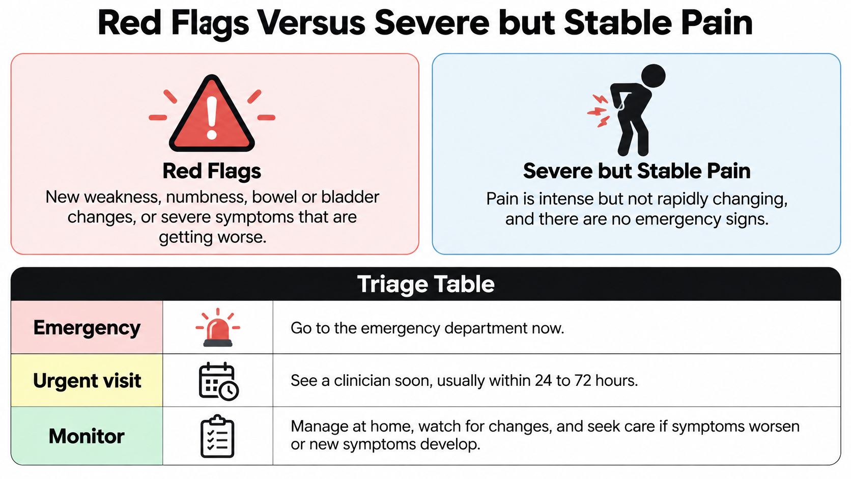
What to do with each warning sign
| Finding | Why it matters | Action |
|---|---|---|
| New trouble urinating, retention, or overflow leaking | Possible cauda equina or severe compression | Emergency evaluation now |
| New loss of bowel control | Same urgent nerve concern | Emergency evaluation now |
| Numbness in the saddle, groin, or genital area | Saddle anesthesia is a classic warning | Emergency evaluation now |
| Both legs weak or numb, or strength dropping fast | Possible progressive neurologic deficit | Emergency evaluation for rapid progression; otherwise same-day urgent assessment |
| One-sided leg or arm pain with numbness or weakness, bladder normal | Can fit nerve-root irritation | Prompt clinical exam, not self-diagnosis |
| Severe one-sided pain but stable strength and normal bathroom control | Often managed first without emergency surgery | Clinical assessment and conservative care |
| Fever, cancer history, unexplained weight loss, or major trauma with spine pain | Possible infection, fracture, or other serious disease | Do not wait for a routine visit |
Saddle sensation means feeling in the area that would contact a bicycle seat or saddle, including the groin, inner thighs, and area around the anus. A new change there isn't an ordinary extension of a sore back.
Urinary retention can also be confusing. It means trouble emptying the bladder, not just needing to urinate more often. Leaking can sometimes occur because an overfull bladder isn't emptying properly.
Stable doesn't mean harmless or easy
Severe but stable pain can still interrupt sleep, prevent work, and make walking difficult. It deserves care rather than being dismissed because emergency signs are absent.
The distinction is about timing and setting, not whether your suffering counts. Emergency services assess possible threats to nerve function; a prompt outpatient examination can evaluate stable symptoms and establish a treatment plan.
Don't wait for an outpatient MRI appointment if an emergency warning sign appears. Likewise, don't assume that normal bathroom control makes rapidly worsening weakness safe to watch at home.
What a Physical Exam Looks For
A useful visit begins with your story, then checks whether your body's responses fit it. The clinician isn't just finding the spot that hurts most.
The NICE briefing paper on low-back pain and sciatica supports combining the symptom pattern with neurologic assessment and using imaging selectively, rather than routinely scanning uncomplicated back pain.
Prepare a clear symptom history
Before the appointment, make brief notes about:
- The route: Where does pain start, and where does it end? Include the buttock, calf, foot, shoulder, hand, or fingers involved.
- The side: Note whether symptoms stay on the same side or affect both limbs.
- The triggers: Describe what happens with coughing, sitting, standing up, bending, or walking.
- The strength change: Record when you first noticed foot drag, difficulty pushing off, dropping objects, or another lost ability.
- The context: Include an injury, previous episodes, and any bladder, bowel, or saddle-sensation changes.
Try a sentence such as: My pain travels from the right buttock into the outer calf, part of my foot feels numb, and my foot has started catching when I walk. That gives the clinician more actionable information than a pain score alone.
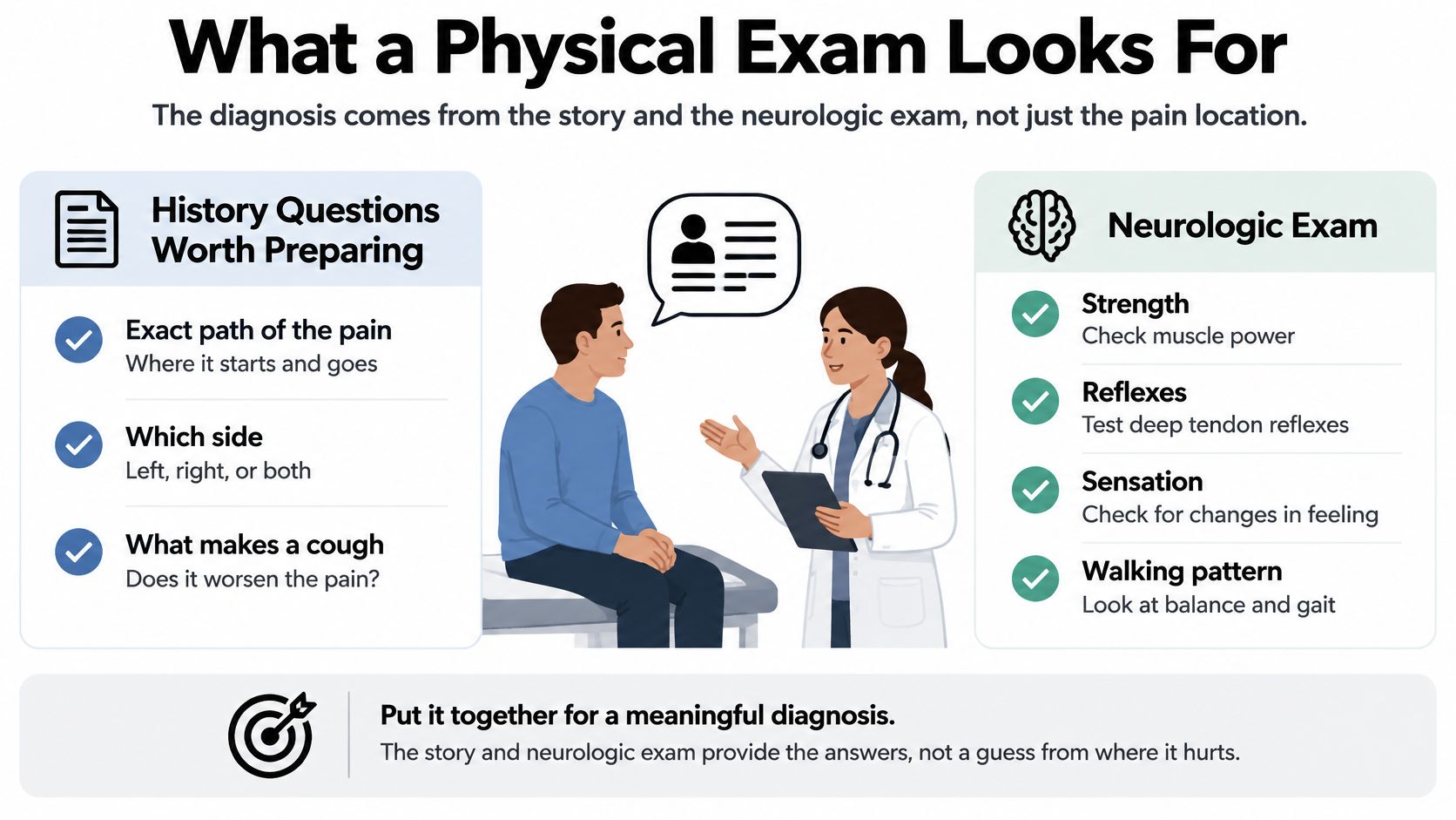
How the clinician compares both sides
The examination looks for agreement between sensation, muscle function, reflexes, and walking. Medical terms such as dermatomal and myotomal refer to skin areas and muscle actions associated with particular nerve roots.
| Check | What the clinician does | Abnormal result suggests |
|---|---|---|
| Dermatomal sensation | Compares light touch and, when appropriate, pin sensation on each leg | A dull strip of skin on one side can fit a nerve root |
| Myotomal strength | Tests heel walking, toe walking, rising on a leg, or grip for neck symptoms | One-sided weakness, such as foot drag or weak grip |
| Reflexes | Compares knee and ankle responses, or arm reflexes for neck symptoms | A quieter response may support nerve involvement |
| Gait | Observes walking for a limp or slapping foot | A possible nerve or muscle deficit |
| Straight-leg raise | Lifts the leg with the knee straight | Reproduction of familiar leg pain can suggest nerve tension |
| Muscle tenderness | Presses the sore area | Tenderness without nerve findings leans toward a non-nerve source |
A straight-leg raise isn't positive merely because the hamstrings feel tight. The clinician considers whether it reproduces your familiar symptoms and whether other findings support the same explanation.
For neck symptoms, controlled positioning may help assess nerve irritation. Don't try forceful neck-compression maneuvers or ask someone at home to perform them.
The result may be a working diagnosis rather than immediate certainty. A documented examination also provides a baseline for deciding whether strength and sensation are improving at follow-up.
Why a Scan Cannot Decide Alone
A spinal disk is a little like a jelly doughnut: it has a tougher outside and a softer center. The comparison helps explain a herniation, but it doesn't tell you whether that structural change is responsible for your symptoms.
The important question is whether the affected nerve matches the painful or weak area. A report describing an abnormal disk is an anatomical description, not a complete diagnosis.
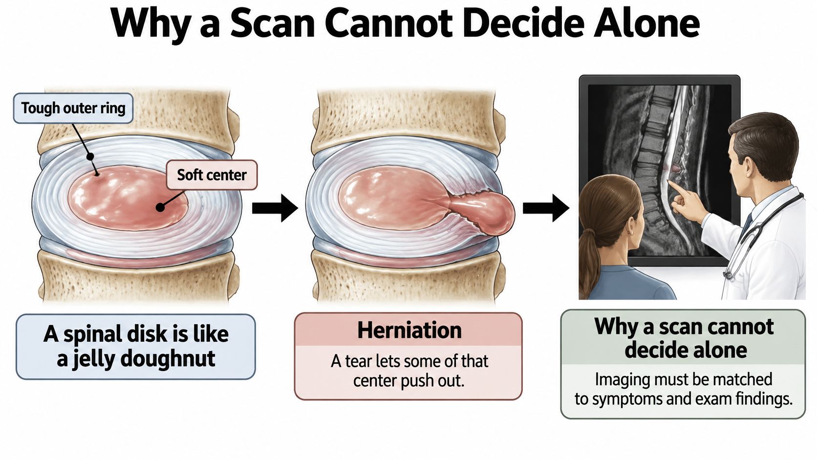
Read the report by level, side, and nerve
The NICE scope document on low-back pain and sciatica describes the relationship between lumbar nerve-root involvement and sciatica, and supports interpreting imaging in its clinical context.
A clinician asks:
- Which level? Does the disk abnormality involve the part of the spine that could explain your symptoms?
- Which side? Does the reported nerve involvement occur on the side where you hurt or feel weak?
- Which findings agree? Do the sensory changes, muscle weakness, and examination support involvement of that nerve?
For example, a right-sided finding that matches right-sided radiating pain and measured weakness is more persuasive than a left-sided bulge in someone whose symptoms are entirely on the right. Even then, the clinician considers other possible causes.
Choose imaging for a decision
MRI shows disks, nerve roots, and other soft tissues without ionizing radiation. Plain X-rays don't show a herniated disk, although they can help evaluate bone and alignment problems.
The practical distinction between these tests is explained further in when you need an MRI and when you need an X-ray. Neither test replaces a hands-on assessment.
The MSD Manual's guide to herniated disks explains why symptom-free disk abnormalities must be interpreted carefully. A scan can reveal a bulge that isn't causing trouble, while severe pain can come from structures or conditions that aren't explained by a disk finding.
A normal MRI doesn't make your pain imaginary. An abnormal MRI doesn't automatically make a procedure necessary.
Imaging becomes more useful when persistent symptoms, progressive deficits, suspected serious disease, or a possible intervention creates a decision the scan can help answer. Waiting on imaging in an uncomplicated case can therefore be a deliberate clinical choice, not a refusal to investigate.
Bring your symptom map and the date weakness began. Those details help the clinician decide what an imaging result would change.
Conservative Care While the Nerve Calms
When warning signs are absent, initial care often focuses on improving movement and function without surgery. Conservative care is active care, not an instruction to stay in bed until every symptom disappears.
A workable plan should fit what you can tolerate today while giving you a clear way to increase activity. It should also explain which changes mean you need reassessment.
Start with manageable movement
During a flare, briefly reduce activities that sharply aggravate symptoms. Then resume walking and ordinary tasks in smaller, tolerable amounts rather than alternating complete inactivity with a large burst of effort.
- Reduce the load: Divide chores, move heavy objects with help, and avoid repeatedly provoking the same radiating pain.
- Keep movement tolerable: Use shorter walks and position changes instead of forcing a distance or posture that consistently worsens symptoms.
- Build capacity gradually: Increase activity according to your response and the clinician's examination, not a fixed deadline.
Physical therapy can include symptom-guided movements, hip and trunk strengthening, mobility work, and practical changes to sitting and lifting. A therapist may look for movements that bring leg symptoms closer toward the back, sometimes called centralization.
That response can help guide exercise selection, but it isn't permission to push through new weakness or spreading numbness. The right exercise is individualized; someone else's sciatica routine may not suit your pattern.
Make pain relief support activity
Manual therapy, targeted stretching, heat, cold, or electrical stimulation may help make movement more manageable. They're supporting tools, not proof that a disk has been pushed back into place, and they don't replace rebuilding strength and tolerance.
Medication choices belong with the prescribing clinician, who can consider your other conditions and medicines. The practical aim is enough symptom control to participate safely in daily life and rehabilitation.
For readers whose symptoms began after childbirth, The Axelrad Clinic's Holistic care for postpartum back discomfort offers related context. Postpartum back discomfort still needs an individual assessment rather than an assumption that every ache is disk-related.
Change the plan when function changes: Increasing weakness needs prompt reassessment, and emergency warning signs need emergency care rather than another exercise session.
Agree on a follow-up point when treatment begins. Don't treat a preset recovery window as permission to wait through deterioration.
If function remains substantially limited despite appropriate care, the clinician may reconsider imaging, medication, an injection, or surgical referral. Those decisions depend on the examination and your progress, not whether the pain was severe at the beginning.
What Recovery Usually Looks Like
Recovery is easier to judge when you track what you can do, not just whether the pain is completely gone. You might first notice that you can finish a grocery trip, sit more comfortably, or get dressed without repeatedly stopping.
Symptoms don't always improve in a straight line. A difficult day doesn't automatically mean a new injury, but a persistent loss of function deserves attention.
Track a few concrete changes
Keep a brief record using the same ordinary activities:
- Pain distribution: Is discomfort staying closer to the back, remaining in the same place, or reaching farther down the limb?
- Sensation: Is the numb area shrinking, unchanged, or spreading?
- Daily capacity: Can you walk farther, sit longer, or complete a familiar task with less interruption?
- Strength: Are you catching your toes, struggling to push off, or dropping things more often?
Don't use repeated maximal lifting as a progress test. Choose safe, repeatable activities that matter in your life and review them with your clinician.
Agree on what success means
A useful target might be walking your usual neighborhood route, returning to a work task, or demonstrating strength closer to the other side. These are more informative goals than merely hoping the back feels normal.
For perspective on duration, how long sciatica pain lasts can help frame the discussion. Your clinician should still base follow-up on your own symptom behavior and examination rather than a general timeline.
If progress stalls, reassessment can clarify whether the exercise dose needs changing or whether another evaluation is appropriate. New weakness or bathroom changes aren't an ordinary setback to exercise through.
After a flare related to lifting or prolonged sitting, adjust the provoking task and discuss how to resume your routine. The aim is a sustainable return to activity, not permanent avoidance of bending, sitting, or work.
Book an Exam Before You Decide
If you're asking, Do I have a herniated disk?, the most useful next step is usually to turn your observations into a clinical examination. You don't need to arrive knowing the diagnosis.
The appointment should help distinguish pain-limited movement from nerve weakness, identify whether the symptom map is consistent, and establish what needs monitoring. That gives you a reasoned next step instead of a decision driven by fear or an isolated phrase in an MRI report.
Bring information that changes decisions
Prepare a short written note rather than trying to remember everything while you're uncomfortable:
- Your symptom map: Mark where pain travels and where sensation feels different.
- Your functional change: Describe what your arm or leg can no longer do reliably.
- Your timeline: Note when symptoms began and when weakness first appeared.
- Your safety information: Report bladder, bowel, or saddle-sensation changes directly.
- Your daily demands: Explain the lifting, driving, sitting, or walking your work and home life require.
If you already have imaging, bring the report so it can be considered alongside the examination. Don't feel obligated to obtain a scan before seeking assessment.
Choose care that fits the finding
For readers in Deerfield Beach and nearby communities, an in-person visit offers a practical alternative to guessing at home. Emergency warning signs belong in emergency care, not in a routine rehabilitation booking.
At MedAmerica Rehab Center, we treat back pain, sciatica, neck pain, headaches, arthritis, and limb pain, and we can develop a pain management and prevention plan suited to your needs. Depending on the service, care may include manual therapy, strengthening exercises, hot or cold packs, electrical stimulation, stretching, or an adjustment.
The first decision is what kind of assessment you need, not which treatment you should buy. Start with the symptoms and function you have today, then use the examination to decide what comes next.
MedAmerica Rehab Center offers individualized physical therapy and chiropractic care for back pain and sciatica, with treatment suited to your needs. If your symptoms are stable and you have no emergency warning signs, visit MedAmerica Rehab Center to arrange an assessment and bring your symptom map, weakness timeline, and questions about imaging.
MedAmerica Rehab · Deerfield Beach
Talk to a clinician about your pain
Physical therapy, chiropractic, acupuncture and shockwave therapy under one roof, since 1995. Same-day appointments are available.
Mon–Thu 8:00 AM – 6:30 PM · Fri 8:00 AM – 3:00 PM